Bewersdorf Laboratory
pan-ExM
pan-Expansion Microscopy (pan-ExM) is a super-resolution technique that offers optical contrast equivalent to EM heavy-metal stains. By physically expanding a biological sample about 20-fold in every dimension, bulk (pan-) staining of proteins or other molecules can now reveal the totality of subcellular organization. Using just a confocal microscope, structures down to ~10-40 nm size are resolved. By further combining pan-stains with antibody labels of specific proteins in multicolor images, images resembling those of correlative light and electron microscopy (CLEM) can be obtained. Our lab is developing this technology for the study of cultured mammalian cells, mouse brain tissue sections, human tissue and other biological samples.
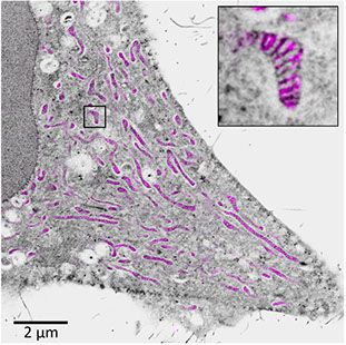
pan-ExM of mitochondria in HeLa cell
