Bewersdorf Laboratory

1
2
3
4
5
STED (‘stimulated emission depletion’) microscopy is a laser scanning fluorescence microscopy method that takes advantage of photophysical properties of the imaged fluorescent probes to create effective scan focus sizes far below the size of the conventional focus of a laser scanning microscope. This way the resolution limit imposed by diffraction can be broken and spatial resolution well below 100 nm can be achieved.
Click the orbs below to learn more...

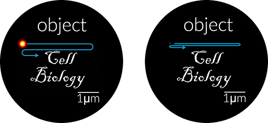
In regular fluorescence microscopy, the fluorophores are excited by a laser beam at a wavelength blue-shifted relative to their emission spectrum (for example at 488 nm for a GFP-like spectrum). Typically, a few nanoseconds after excitation, the fluorophores relax spontaneously into their electronic ground states by emitting fluorescence (with the major emission contribution between 500 and 550 nm). In STED microscopy a second laser beam with a wavelength tuned to the red tail of the fluorophores’ emission spectrum is added (at approximately 590 nm for a GFP-like spectrum ). The STED laser beam can stimulate emission of light from the fluorophore to force the molecule down to its electronic ground state before spontaneous fluorescence can occur. Thus, fluorescence can be ‘switched off’ by the STED laser beam.
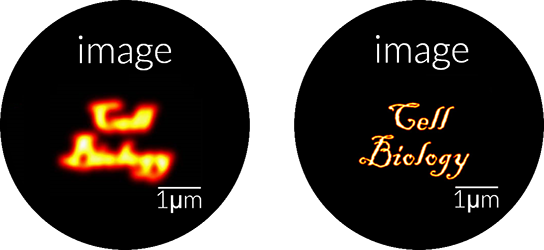
To achieve resolution enhancement, the STED beam profile is modified to produce a ring-shaped intensity profile centered on the excitation focus with a central minimum of ideally zero intensity. This way fluorescence is switched off in the outskirts of the excitation focus. The fluorescence emission volume is narrowed by raising the STED laser power, saturating the depletion process this way: by increasing the power, a larger and larger area of the ring profile reaches intensity values sufficient to deplete excited fluorophores. The central spot of ‘zero’ intensity, however, remains at ‘zero’. This results in a smaller and smaller area in which fluorescence can occur. Try increasing the STED laser power by clicking the +sign.
Confocal
STED
Scanning a sample with this reduced effective focus volume creates images of greatly enhanced spatial resolution well below 100 nm.

Our current optical development projects supplement the super-resolution capabilities of STED microscopy with adaptive optics, two-photon excitation, and 4Pi optics. These innovations allow fast live-cell and multicolor imaging as well as 3D imaging deep in living animals.
Moreover, our state-of-the-art 4Pi-STED system achieves tissue imaging with sub-50 nm resolution in 3D. Please visit our publications page to see our latest developments.